In the human body, the femur (thighbone) is the strongest and the longest bone. Since the femur is strong, it generally takes a high force like motor vehicle collisions to break it. So, fractures along the length of the femur are femur shaft fractures.
The straight and long part of the femur is its shaft (femoral shaft). If a break occurs anywhere along this bone length, it is a femur shaft fracture. Femoral shaft fracture usually requires surgical treatment.
Types of Femur Shaft Fractures
Generally, femur fractures can be classified on the basis of force applied, location and pattern of the fracture, and nature of the injury.
1. On the basis of force causing the break:
If the pieces of the bone line up correctly, it is a stable fracture, or if it is out of alignment then it is a displaced fracture. If the skin around the fracture is intact, it is a closed fracture or if the bone ruptures the skin, it is an open fracture.
2. On the basis of location:
The femoral shaft has three parts: distal, middle, and proximal and anyone can break it.
3. On the basis of the pattern of the fracture:
For e.g. the bone can break in different directions, such as crosswise, in the middle, or lengthwise.
4. On the basis of injury:
If due to the injury, skin, and muscle over the bone are then the most common types of femur shaft fractures are transverse, oblique, and spiral. If the break is in a straight horizontal line going across the femoral shaft, it is a transverse fracture. When the fracture has an angled line across the shaft, it is an oblique fracture. And if the fracture line encircles the shaft just like the stripes on a candy cane, it is a spiral fracture. This type of fracture occurs by a twisting force on the thigh.
Comminuted fracture – When the bone breaks into three or more pieces. Usually, the number of bone fragments corresponds with the amount of force required to break the bone.
Open fracture – When the bone fragments stick out through the skin or a wound penetrates down to the broken bone, then it is termed as a compound or open fracture. This type of fracture includes severe damage to the surrounding muscles, ligaments, and tendons. These fractures are more complicated and have a high risk of infection. Moreover, the healing time for these fractures is also more.
What Are the Causes?
Generally, in young people, femur shaft fractures occur because of some type of high-energy collision like a vehicle or motorcycle crash, collision with a car while walking falls from heights, and gunshot wounds. These are some of the common reasons. But in the case of aged patients, a lower-force incident like a fall from standing may be potential enough to cause a femur shaft fracture.
Femur Shaft Fracture Symptoms
The femur shaft fracture symptoms include immediate severe pain, inability in putting weight on the injured leg, and even it may look deformed i.e. shorter than the other leg and not straight.
Examination of Fractures in the Femoral Shaft
A thorough examination is a must to diagnose fractures in the femoral shaft. Let us have a look at the same:
Medical History and Physical Examination
The doctor must know the particular details regarding the injury. For e.g. mechanism of injury. Because if it is a result of a car accident, the doctor must know the speed of the car, whether the patient was a driver or a passenger, whether he/she was wearing a seat belt or not, and whether the airbags went off or not. These details will greatly help a doctor to determine the reason and mechanism of injury.
A doctor should know about the patient’s health conditions, such as high blood pressure, diabetes, asthma, or allergies, and if he/she is taking any medication or using tobacco products.
After a comprehensive understanding of the injury and medical history, a doctor needs to do a careful examination and assessment of the overall condition. While a physical examination, a doctor should check:
- A deformity of the thigh or leg (an unusual angle, shortening of the leg, or twisting)
- Breaks in the skin
- Bruises
- Bony pieces pushing on the skin if any
After examining these conditions, a doctor must feel for pulses. If the patient is awake, the doctor will examine sensation, and movement in the leg and foot. Then the doctor should examine the patient’s leg, thigh, the tightness of the skin, and muscles around the thigh and foot and then find out the abnormalities.
Femur Shaft Fracture Treatment
The treatment of femur shaft fracture depends on the severity and the type of fracture. However, most fractures of the femur shaft will require surgery. Let us see available treatment options:
Non-surgical Treatment
However, in most cases, femur shaft fractures need surgical treatment. But in some cases, non-surgical treatment is enough. In the case of very young children, the doctor will apply a cast.
Timing of surgery- In the case of surgical treatment, femur fractures should be fixed within 24 to 48 hours. Fixation can be delayed so long as other life-threatening injuries or unstable medical conditions are stabilized. Open fractures should be treated with antibiotics as soon as the patient is admitted to the hospital so that the risk of infection can be minimized. The surgeon will clean the open wounds, bone, and tissues.
Prior to surgery, a doctor can place the leg either in a long-leg splint or in traction as emergency management of the fracture. Skeletal traction is a pulley system of weights and counterweights that holds the broken pieces of bone together. It keeps the leg straight and also helps in relieving the pain. It keeps the broken bones as aligned as possible and maintains the proper length of the leg.
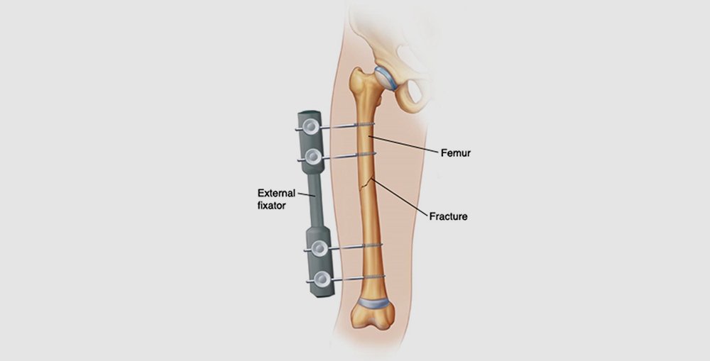
External fixation
For external fixation, metal pins or screws are placed into the bone above and below the fracture site to hold the bone inadequate position. These pins and screws are attached to a bar outside the skin thus, this device is a kind of stabilizing frame for the fractured bone.
Generally, external fixation is a temporary treatment for femur fractures. External fixators are feasible in case of multiple injuries and if the patient’s condition is not suitable for a longer surgery for fracture fixation. An external fixator can provide good, temporary stability unless the patient’s condition is suitable enough for definitive surgery. Sometimes an external fixator can be left on until the femur is fully healed completely but it requires rarely.
Intramedullary Nailing
Intramedullary nailing is the common procedure for treating femur shaft fractures. Usually, intramedullary nails are of titanium. They are available in different lengths and diameters to fit most femur bones. In this procedure, a metal rod designed specifically is inserted into the femur canal. Intramedullary nails can be inserted into the canal either at the hip or the knee. The rod passes across the fracture such that it can keep it in position. To hold the leg in the correct alignment, the surgeon will place the screws below the fracture.
Plates and Screws
Plates and screws are usually applied when intramedullary nailing is not possible as in the case of fractures that extend into either the hip or the joints of the knee. During surgery, the surgeon will first reduce the fracture to the normal alignment. Bone fragments are held together with screws and metal plates are attached to the outer surface of the bone. A wide range of orthopedic implants and tools such as the tibia and femur interlocking systems are made available by orthopedic implant manufacturers.
Recovery
Complete healing of most femur shaft fractures may take 3 to 6 months. In some cases, healing time may be longer, particularly when the fracture is open or broken into many pieces or if the patient consumes tobacco products.
Pain Management
A doctor and nurses can work to reduce the pain, which can also facilitate a speedy recovery. After an injury or operation, pain is a natural part of the healing process. For short-term relief, medications will help after the injury and surgery. The medicines available for the management of reducing pain include acetaminophen, gabapentinoid, nonsteroidal anti-inflammatory drugs (NSAIDs), muscle relaxants, opioids, and other medications for topical pain. A doctor may prescribe a combination of medicines for pain relief, as well as to minimize the need for opioids. However, these medications may have side effects that may affect a patient’s ability to drive and perform other activities. A doctor can discuss with the patient the side effects of these medications.
Since opioids are narcotics so can be addictive. Overdose and reliance on opioids are also serious public health issues in the U.S. So, doctors’ advice is a must before taking these medicines. One must stop taking opioids once the pain starts to improve. If pain persists even after a few days of treatment, the patient should consult the doctor.
Weight-bearing
To avoid any complications, it is important to follow the doctor’s instructions for weight-bearing on injured legs. In some cases, doctors can encourage leg motion in the early recovery period. But in other cases, the doctor will allow weight-bearing (as much as possible on the leg) right after surgery. Though a patient may be unable to put full weight on the leg unless the healing process has started. The patient may need crutches or a walker for support once he starts walking.
Physical Therapy
Since a patient tends to lose muscle strength in the injured area, exercises during the healing process are very significant. Physical therapy facilitates the restoration of normal muscle strength, flexibility, and joint motion. It also helps in the management of pain post-surgery.
A physical therapist can start a patient’s physiotherapy when the patient is in the hospital. The therapist will teach the use of crutches or a walker to the patient.
Femur Shaft Fracture Complications
Femur shaft fractures may result in further injury and complications which are as follows:
- As the ends of broken bones are sharp so they may tear or cut the surrounding nerves and blood vessels.
- A painful condition namely acute compartment syndrome may occur. This syndrome develops when extreme pressure within the muscles happens to be at a dangerous level, this may reduce the flow of blood which further hinders the supply of oxygen and nourishment to nerve and muscle cells. If the pressure is not relieved quickly, it may result in permanent disability or a surgical emergency. Then to release the pressure, the surgeon will make incisions in the skin and the muscle coverings.
- There may be a risk of infection in open fractures even after proper surgical cleaning. Since the bone is exposed to the external environment in open fractures. Treatment of this infection may be difficult and may require further surgeries and long-term use of antibiotics.
- Sometimes in femur shaft fractures, the ligaments around the knee can also injure. If there is knee pain even after surgery, the doctor must consider it.
Complications from Surgery
Complications of surgery usually involve:
- Infection, blood clots, injury to nerves, and Fat embolism.
- When bone marrow enters the bloodstream and can travel to the lungs. The fracture itself may cause this.
- Malalignment or the incorrect positioning of the broken bone fragments.
- Non-union or delayed union results when the fracture does not heal or heals slower than usual.
- Hardware irritation occurs when the overlying muscles and tendons irritate due to the end of the nail or the screw.